Heavy Metals: A Missing Link in Infertility?
Infertility affects one in eight couples, with the disorder stemming from both men and women at about the same rate. While clinics and doctors focus on hormones, age, and other biological factors, a well established but largely overlooked factor is the role heavy metal toxicity plays in male and female infertility.
In this article we review the current research and then give you direction in how to protect your fertility from heavy metal toxicity.
What you’ll learn in this article:
- Although infertility is multi-faceted, the role of heavy metal toxicity is well established and often overlooked.
- Heavy metal toxicity has about the same impact on fertility in both men and women.
- Although heavy metal toxicity and fertility still need more study, the research that has been done thus far provides solid evidence on the links between heavy metal toxicity and infertility.
- In women, heavy metal toxicity impacts follicular health, egg development, egg implantation, hormone balance, fetal development, genetic expression, and other aspects of female reproduction.
- In men, heavy metal toxicity impacts testicular health, development and function of the sperm and semen, the ability of the sperm to penetrate the egg, genetic expression, and hormone balance.
- Although our environment is inundated with heavy metal toxicity from industrial use, fortunately there are things we can do to remove these toxins. .
Infertility is a multi-faceted disorder that affects one in eight couples trying to get pregnant today, with the disorder stemming about equally from both the female and male partners. While hormonal and reproductive issues are most often cited as the causes of infertility, there is one glaring but overlooked factor that affects us all: Heavy metal toxicity.
Our environment is heavily laden with toxins and heavy metals since the rise of the industrial revolution. Heavy metals are in our air, soil, water, foods, and the everyday products we use. We are also exposed through cigarette smoke, supplements and alcoholic beverages (1).
Heavy metals pose health risks because our bodies cannot metabolize them for one reason or another and because they persist so long in the environment and in our bodies. They affect every different metabolic function in our body, including many different mechanisms required to successfully procreate.
Sadly, manufacturers are for the most part not required to test the safety of chemicals and heavy metals in foods, food packaging and personal care products prior to use, so they have been allowed to infiltrate our environment for decades without first being tested for human safety.
However, as research on the effects of heavy metals on human physiology grow, so does our knowledge on how they affect the many intricacies of reproduction in both women and men. The news is not good — the tens of thousands of toxins and the abundance of heavy metals in our environment is negatively impacting our health and our ability to have children.
The good news is, it is possible to help buffer the body from exposure to heavy metals, detox them from the system, and create more resilient health. You can learn more about this here and I’ll also talk a bit more about it at the end of the article.
However, in this article, we are going to take a journey through the current research on the links between heavy metals and infertility.
I am going to start with an overview of research on the impact of various heavy metals on infertility, and then delve more deeply into a handful of heavy metals that have been studied more extensively.
An Overview of Heavy Metals That Impact Fertility
Heavy metals are used in many industries and research has clearly established their threat to human health. The danger of heavy metals in our environment comes less from acute, one-time exposures and more from accumulation in body tissue over time due to their prevalence in our everyday world and the body’s inability to detoxify them adequately (2).
Not all metals are bad. In fact, some are necessary for human health at biologically appropriate levels. These include copper, iron and nickel, but in excess are toxic to the body. However, other metals have been shown to be harmful to human health and are over abundant in our environment due to their industrial uses. These include arsenic, cadmium, mercury, lead, aluminum, uranium, and more.
Unfortunately, very few broad studies that examine trends in human populations exist when it comes to heavy metals and infertility.
However, we can look at numerous studies that explore the effects of heavy metals on various aspects of female and male reproduction.
Lead. For example, some studies show links between elevated blood levels of lead and infertility in women. Blood and serum lead levels were significantly higher in women with pre-eclampsia than in women without pre-eclampsia (3, 4).
Mercury. Studies also show reduced fertility among dental health care workers who performed procedures that exposed them to mercury (5).
Additionally, a 2006 study showed a significantly negative association between blood mercury levels and the ability to carry a pregnancy to term in first-time pregnant mothers (6).
Maternal elevated blood mercury levels, cord blood mercury levels, and maternal urinary lead levels were also significantly associated with low birth weight (7, 8, 9).
Arsenic. Some animal studies have shown results consistent with human population-based studies (also known as epidemiological studies) when it comes to arsenic. Rat studies showed sodium arsenate, a more toxic form of arsenic, caused constant diestrus, or lack of fertility (10, 11).
High levels of arsenic in drinking water and blood lead levels were significantly associated with increased odds of spontaneous abortion (12, 13).
Elevated maternal blood, hair, and urinary arsenic levels have been associated with low birth-weight or small infants (14, 15,16).
The Link Between Heavy Metals and Infertility is Consistent In the Research
Additionally, high arsenic levels in drinking water, elevated urinary levels of arsenic (17), maternal blood lead levels, placental lead levels, maternal environmental mercury exposure, maternal hair mercury levels, and cord blood mercury levels were significantly associated with shortened gestation or preterm birth (18, 19, 20, 21, 22, 23, 24).
And yet several other studies report no association between any of these markers and low infant birth weight (25, 26, 27, 28, 29, 30, 31, 32, 33, 34, 35).
Although there are conflicting study results, overall we cannot deny that heavy metal exposure has been shown to interfere with female and male reproduction in both experimental and epidemiological studies.
When considering the conflicting results several factors must be taken into consideration: Differing sample sizes, use of different tissue types to measure heavy metals, or genetic variation between studied populations.
In fact, some researchers conclude that the link between heavy metals and infertility is consistent across different statistical modeling strategies described (36, 37, 38).
The Effects of Heavy Metals on the Ovaries
Studies on the association between heavy metals and ovarian health are conducted mainly to support fertility technology, namely in-vitro fertilization (IVF). However, they also give us insights into the links between heavy metals and infertility.
For instance, one study reported that women undergoing IVF treatments in Taranto, Italy, an industrial area known for its heavy metal contamination, have significantly elevated levels of heavy metals in their follicular fluid, including lead, compared to women undergoing treatment in less polluted areas (39).
Further studies showed high mercury levels in hair samples was correlated with lower yield of oocytes, or eggs, in the follicles (40), and that hair concentrations of mercury above EPA standards of 1ppm is linked to lower egg yields than in those with lower hair levels of mercury (41).
Another study found a relationship between elevated sodium arsenate in drinking water and poor ovarian and follicular health in rats (42), while another mouse model showed that topical skin creams containing mercury caused a significant accumulation of mercury in the ovaries (43).
The Effects of Heavy Metals on the Uterus
Although limited data exists on the associations between heavy metals and uterine health, we can look at a few studies. For instance, elevated urinary levels of lead and blood levels of mercury were significantly associated with fibroids in 99 women compared to a control group of 374 women (44).
Rat studies have shown that exposure to sodium arsenite through drinking water resulted in a smaller uterine size and poor uterine development (45), and impacted genes involved in uterine development (46).
Increased cadmium levels have also been associated with lower egg fertilization (47) and implantation rates (48).
The Effects of Heavy Metals on Sperm and Male Reproduction
We tend to associate infertility with women, but male infertility rates almost equal that of females.
For instance, research has found that abnormal sperm activity is associated with elevated levels of a wide range of heavy metals compared to controls, including aluminum, arsenic, antimony, lead, chromium, tin, nickel, tungsten, and uranium (49, 50, 51).
Arsenic and cadmium are well-documented as being toxic to the male reproductive system, as they reduce sperm motility in a linear dose-dependent manner. In other words, the higher the body burden of these heavy metals the larger the impact on sperm health (52, 53, 54, 55, 56).
Researchers have found heavy metals such as cadmium and arsenic suppress sperm motility by dysregulating the spermatozoa antioxidant system (57, 58).
The male reproductive system is highly dependent on a healthy antioxidant system and oxidative damage can immobilize sperm by inhibiting enzymes in the testes necessary for the development of healthy sperm cells (59, 60).
Additional studies have found that elevated levels of lead may lead to abnormal sperm and sperm development (61). Moreover, exposure to high levels of arsenic and copper can damage sperm DNA integrity (62).
The Research Linking Common Heavy Metals and Infertility
Although the effects of all heavy metals on human health hasn’t been studied, and although the impacts of heavy metals on fertility still requires further study, we do know a number of metals are known to be toxic and exist in high enough levels in our environment to be a risk to human health and fertility.
We also know that while discrepancies in studies exist, ample research nevertheless demonstrates negative impacts on female and male reproductive physiology in a way that raises the risk of infertility (63).
In these next sections I’ll summarize the most abundant research showing the link between infertility and cadmium, arsenic, mercury, lead, aluminum and uranium.
CADMIUM
Cadmium and infertility
In a literature search for associations between infertility and heavy metals, cadmium produces many results. And with good reason. Cadmium is one of the most toxic heavy metals, has no known beneficial biological function, and poses a significant public health hazard.
Cadmium is abundant in our soil, air, water, and food. Coal burning releases cadmium and other metals into the air where it ends up in the ocean and gets into our food supply via fish and shellfish. It is found in cigarette and marijauna (even medical marijauna), used in rechargeable batteries (think improper battery disposal leaching into soil), as a pigment in paint, and in plastic production.
It is also released into the environment through mining, smelting, waste incineration, and electroplating of consumer products. It is extruded during the processing of other metals such as zinc, copper, and lead. It can also permeate the environment through wildfires and volcanic activity.
However, in the US, the most common sources of cadmium exposure are ingesting it through contaminated foods and inhaling it through smoking and second-hand smoke. Food, even organic, uses fertilizers. Many phosphate fertilizers are from Morocco and are contaminated with cadmium. Some fertilizers come from wastewater treatment solids that have high levels of cadmium. Even organic food can be fertilized with this wastewater sludge – the result of removing the solids left over after water treatment.
Food sources most contaminated with cadmium are leafy greens, potatoes, grains, peanuts, soybeans, sunflower seeds, large migratory fish, shellfish, organ meats, rice, wheat, and celery root (64, 65).
For smokers, their blood concentration levels of cadmium — 1.58 µg/L — far surpass that of non-smokers at only 0.38 µg/L (66).
Cadmium accumulates in the liver, kidneys, testes, ovaries, and the placenta (67).
Cadmium toxicity in the placenta has been shown to cause cell damage, genetic alterations, and a decline in the production of the pregnancy hormones progesterone and human chorionic gonadotropin (HCG) that is needed to create the placenta to feed a fetus (68, 69). Cadmium damages cells and tissue by promoting the production of reactive oxidative species that cause free radicals and oxidative stress (70).
Cadmium even impacts the outcomes of IVF. A study of infertile couples assessed whether exposure to lead, mercury, and cadmium prior to conception affects in vitro fertilization (IVF) outcomes. Researchers found that cadmium toxicity was associated with lower fertilization of the egg by the sperm (71) and lower egg implantation rates (72).
The Effects of Cadmium on the Developing Fetus
Cadmium impacts fetal development by competing with zinc and copper in the fetal liver, brain, kidney, and intestines (73). By taking the place of these essential minerals in the developing fetus, cadmium toxicity leads to abnormal growth and development, which may persist into adulthood (74).
Exposure to cadmium during pregnancy has been associated with low birth weight, smaller head circumference, and premature birth, affecting infant females more than males (75, 76, 77, 78, 79, 80). These are the very same outcomes seen in children whose mother’s smoke during pregnancy or who quit right before getting pregnant, yet are still full of fetus-toxic cadmium. But the cause is really the cadmium ingested from cigarette smoking.
While the reason for this imbalance in cadmium affecting females more than males is not fully understood, it has been shown that cadmium impacts fetal DNA in female infants more so than males (81).
Cadmium also causes changes in the expression of placental DNA (82). It does this by inhibiting the process whereby cells divide and the DNA is copied incorrectly, causing genetic mutations. A survey study of preconception cadmium exposure showed it impacted gene expression in the umbilical cord (83), while other studies have also observed genetic effects related to cell death (84). Elevated levels of cadmium in cord blood was associated with emotional and cognitive problems in boys ages 7 and 8, but not girls (85).
Cadmium is also an endocrine disruptor, meaning it can mimic or inhibit the body’s own estrogen. This causes hormonal imbalances like estrogen dominance or low estrogen in both females and males (86).
Cadmium Exposure and Male Infertility
In men, studies have shown cadmium can decrease testosterone, reduce sperm concentration, and decrease fertilization capacity (87, 88). What’s worse is that this damage persists and can be seen in subsequent generations due to DNA damage.
Other studies have shown that cadmium damages semen, decreases sperm count, and impacts sperm morphology, sperm motility, and sperm viability (89, 90).
Furthermore, cadmium has been shown to cause changes in sperm development, decrease semen quality, damage areas of the testes, and inhibit male hormone function (91). It’s believed oxidative stress from cadmium exposure damages male reproductive tissues and cells and interferes with hormonal signaling pathways (92, 93).
Numerous observational studies have also demonstrated a link between cadmium in both blood and seminal fluid and low sperm viability and motility (94, 95, 96, 97).
In animal studies, cadmium exposure has been shown to cause enormous damage to mature sperm. More specifically, cadmium was shown to be toxic to sperm development, which led to decreased sperm motility and concentration (98, 99, 100).
Because it competes with calcium, cadmium is believed to damage cell membranes and cell structure, thus interfering with processes necessary for the sperm to penetrate the egg (101). The unique structure of sperm makes it dependent on calcium for proper development and function, which may help explain one reason why sperm are sensitive to cadmium.
In general, animal studies have shown cadmium exposure impairs testicular tissue, sperm motility, and the ability of the sperm to penetrate the egg.
A very recent study showed the same effects on fish as well. Research of a South American freshwater fish has shown that cadmium toxicity impaired sperm quality, increased larval mortality, and caused physiological changes in the early stages of development, thus reducing the fertility rate of this vulnerable fish species (102). I presume a similar effect across other species of fish as well.
Although the average man only takes in a small amount of cadmium on a daily basis, the problem is that it accumulates in tissues and can linger 20-30 years in body tissue (103). It accumulates mostly in the liver, kidney and testes (104, 105, 106, 107).
I have witnessed this over the years in testing thousands of clients for heavy metals. Even if someone quit smoking decades prior or were exposed to secondhand smoke as a child, high levels of cadmium still appear on their heavy metals tests years later. It always surprises me, but this result is consistent.
Cadmium Exposure and Female Infertility
Cadmium has also been shown to negatively impact female fertility. It has been well-documented that cadmium exposure negatively affects the maturation of the egg, its fertilization, and embryo development (108).
Cadmium may interfere with oocyte (egg) maturation, fertilization, embryo development, and fertility by impacting cell division and structure, causing oxidative stress and cell and tissue damage, and modifying genetic expressions. It has been found that as cadmium exposure increases, so does infertility (109, 110).
Cell culture studies showed that sperm incubated in a cadmium-enriched medium negatively impacted fertilization and embryonic development compared to controls (111) and caused a decrease in implantation rates (112). However, while the fertilization rate decreased as cadmium exposure increased, eggs fertilized with these sperm still retained their full-term developmental potential after being transferred (113).
One study showed that cadmium exposure decreased the number of ovulated eggs and impaired cell division and maturation rate (114). Additionally, this study concluded that cadmium exposure affected the ovarian follicle microenvironment, which then continually impaired the ability of eggs to mature (115). Embryonic development after fertilization was also impaired (116).
Acute exposure to cadmium is also known to affect the hypothalamic-pituitary-ovarian axis, or the communication between the brain and the female reproductive organs that helps regulate hormone levels. This resulted in a decrease of the reproductive hormones follicle-stimulating hormone (FSH), luteinizing hormone (LH), and basal progesterone levels, which are responsible for ovulation (117, 118, 119).
Another study showed that cadmium exposure reduced the number of ovulated eggs by approximately 20 percent, and impaired egg maturation necessary for it to develop into a fetus (120).
More Heavy Metals That Impact Fertility
ARSENIC
Arsenic and infertility
Arsenic is found naturally in the earth’s crust; however, we are coping with high levels in our environment due to pollution from manufacturing, water contamination and in our food supply, namely chicken, eggs and rice.
Arsenic is used in wood preservatives, pesticides, mining, smelting of ores, and coal-fired power plants. We are exposed to arsenic through contaminated water, food, and air. Exposure typically occurs through ingesting contaminated food or water or through inhalation.
The EPA limit for arsenic in drinking water is 10 parts per billion and an OSHA permissible exposure limit over 8 hours is 10 µg/m3 for airborne arsenic (121). But exposure to these smaller amounts of arsenic over decades accumulates in the tissues leading to low level chronic symptoms.
One of the most common sources of arsenic toxicity comes from water. Some areas have naturally higher levels of arsenic in the soil and groundwater, resulting in an increased exposure for residents in that area (122). And arsenic is a common well water contaminant.
Arsenic impacts female fertility by decreasing the likelihood of sperm being able to fertilize the egg (123). Arsenic can also negatively impact sperm activity by inducing cell death, changing the structure of sperm, decreasing sperm count, and impairing sperm development (124, 125).
Animal studies have shown an overall decrease in fertility in animals exposed to arsenic (126).
A 2012 study found that men with unexplained infertility, which account for about 15 percent of men with infertility, exhibited higher levels of arsenic (127, 128, 129).
Both concentration and toxicity play roles in arsenic toxicity and its impact on fertility, and levels of arsenic in infertile males has been significantly higher than in fertile controls in some studies (130). It is believed this is because exposure to elevated levels of arsenic induces serious oxidative stress and inhibits the body’s ability to metabolize and detoxify arsenic (131).
MERCURY
Mercury and Infertility
Mercury is released into our environment all over the planet through coal burning, waste incineration, and other industrial and manufacturing processes (132).
From these sources it then contaminates soil, air, and water. Released into the air from coal burning and manufacturing, mercury settles into the ocean and bioaccumulates in fish, especially large fish such as tuna, swordfish, shark, tilefish, mackerel, marlin, orange roughy, and cod. It’s also found in lobster and other shellfish (133).
Consuming fish with high levels of mercury is believed to be the primary way people expose themselves to mercury (134). Second would be mercury amalgam fillings. However, mercury is also breathed in the air and found in topical skin creams (skin lighteners), broken thermometers, 400 medications as a preservative, fluorescent lamp manufacturing and exposure can also occur through inhalation of polluted air (135).
Mercury occurs in several forms, and may be found in thermometers, dental mercury amalgam fillings, batteries, antiseptic creams, and skin lightening creams. It is also released into the environment from mining ore deposits, burning coal, and via manufacturing plants, although it also be released through volcanic activity.
Human exposure most often occurs from eating fish or shellfish contaminated with methylmercury, breathing mercury vapors, dental work and medical treatments, and occupational skin exposure. The EPA has set a limit of 2 parts per billion of mercury in drinking water and 1 part per million of methylmercury in seafood (136, 137).
I don’t advocate for avoiding fish while healthy, pregnant or trying to get pregnant simply because it contains mercury. ALL food, whether it is animal protein or vegetables, has toxic metal contamination. I advocate that people are simply aware of what they are ingesting, avoiding sources of clear high metal contamination (i.e. big eye tuna). And doing sensible detox on a regular basis – for life.
Exposure to high levels of mercury can permanently damage the brain and kidneys and impair fetal development. A study of women undergoing IVF showed that high concentrations of mercury in hair samples was correlated with a lower likelihood of egg maturation (138).
Another study of women in Japan showed that infertile women had higher mercury levels and lower levels of selenium, while fertile women had lower levels of mercury and higher levels of selenium (139).
These results suggest that mercury may reduce fertility and that selenium may offset the toxic effects of mercury to the reproductive system (140). Selenium aids in the removal of mercury from the body.
Research has also shown associations between elevated levels of mercury in hair samples and impacts on sperm concentration, total sperm count, and sperm motility in men and in women poor egg yields and follicular health (141, 142) and shortened gestation or preterm birth (143, 144, 145, 146, 147, 148).
Mercury exposure has also been linked with damage to the brain, heart, kidneys, lungs, and immune system in humans of all ages. Additionally, mercury is insidious in that when one has mercury toxicity it reduces the body’s ability to detox, metabolize and release all other toxins and heavy metals by impacting detox methylation pathways and poisoning the body’s ability to produce glutathione.
LEAD
Lead and Infertility
Lead was primarily introduced into our air, water, and soil through leaded gasoline emissions, which is still happening in developing countries (149). Exposure can also occur through drinking water from lead pipes, and ingestion or inhalation of dust and flakes of lead-based paints, which can occur in any home built prior to 1978 (150, 151).
Before 1978, lead was used in gas, paint, and water pipes so people living in older homes are at high risk for lead poisoning, especially during remodeling and renovation (152). You absolutely do not want to sand and repaint a home while living in it, especially if you are pregnant or have children. This can lead to irreparable brain damage in a fetus or child.
Lead can be found in iron ore deposits worldwide and is released into the environment through mining, smelting of ore, manufacturing and application of pesticides, and waste incineration (153).
Working in a battery factory or jewelry making, living in an area without lead regulations, and using glazed cookware and dishware (especially vintage) that have high lead levels are other common sources of exposure. Lead is also found in children’s toys, jewelry, storage batteries, leaded crystal, dark hair dyes and ammunition (think war zones).
Herbal remedies or cosmetics imported from countries with no lead regulations are a source of lead (154). However, even when there are lead regulations there is little oversight. Lipstick and mascara commonly contain lead, even in products sold in the US.
Elevated blood serum levels of lead are associated with gestational hypertension (high blood pressure), preterm delivery, low birth weight, miscarriage, birth defects, and abnormal placental development (155).
One study found that couples in which the male partner had elevated blood levels of lead, they had a 15 percent reduction in the ability to conceive (156). Lead is believed to impact semen quality by decreasing sperm count, motility, and viability, by reducing DNA integrity, and by increasing the likelihood of structural abnormalities in sperm (157, 158, 159).
A study of IVF patient couples found a 47 percent decrease in total mobile sperm count when lead was noted in seminal plasma, the fluid portion of semen (160).
Animal studies have shown that after ingesting water treated with high concentrations of lead, this resulted in a decrease in overall fertility, particularly in relation to sperm motility, structural changes in the sperm and testes, and through fragmenting DNA (161).
It is statistically impossible to not have lead in your body. It is so pervasive in our environment from leaded gasoline emissions and industry that lead has infiltrated most soil where food is grown. And we ingest it in the air, food and water as a result.
ALUMINUM
Aluminum and Infertility
Aluminum is the most abundant metal in the earth’s crust, and can be found naturally in the soil, water and air (162). It is statistically impossible to not have aluminum in your body given it’s prevalence on the planet and its continuous release into the environment from manufacturing.
Aluminum has a vast number of industrial and manufacturing uses to make antiperspirants, food containers, airplanes, roofing, aluminum foil, explosives, antacids, astringents, antidepressants, food additives, an anticaking agent in sugar, salt and flour and numerous other consumer products.
Aluminum is released into the air and the environment through mining, the production of aluminum metals and aluminum compounds, and from coal-fired power plants. Aluminum exposure also comes through the consumption of fresh fruits, vegetables, meat due to environmental exposure (163).
Animal studies have shown that exposure to aluminum doses as low as 1.5 mg/kg were associated with a decrease in sperm count, sperm production, and sperm motility. Aluminum is also associated with testicular inflammation (164). Studies of IVF patients found men with a low sperm count also showed an increase in aluminum concentration (165).
URANIUM
Uranium and Infertility
Depleted uranium poses clear threats to human health and fertility (166).
Uranium is a naturally occurring radioactive element that enters the body through drinking water, diet, inhalation, and skin contact. Many don’t realize that they are showering daily or drinking water that contains uranium that builds up over time.
Uranium is a common water contaminant in city and well water in the southwestern United States. In fact, when scientists from the University of Nebraska took some 275,000 groundwater samples for evaluation, they discovered that many Americans live about a kilometer from wells that are uranium-polluted[180]. What’s more, a recent study conducted by U.S. researchers found that almost 2 million people in California and the Midwest live on aquifer sites which have up to 180 times the safe level of uranium[180].
Depleted uranium is a man-made by-product of the uranium enrichment process used in nuclear power reactors and military weaponry. While depleted uranium is less radioactive, it is nevertheless very toxic and its use in war zones has caused it to become a significant public health concern (167).
When measuring uranium in the body, higher levels are found in seminal fluid than in the blood. Uranium exposure has been significantly associated with decreased sperm motility, viability, or normal structure (168).
Experimental studies on rodents have demonstrated a clear association between depleted uranium, testicular atrophy, and reduced sperm development (169).
CHROMIUM
Chromium and Infertility
Chromium is found naturally throughout the natural environment. However, there are a few forms of chromium, some more toxic than others. Chromium(0) is a metal and is used to make steel. Chromium(III) is used to tan leather, preserve wood, and in dyes, pigments, and chrome plating.
Chromium is released into the environment from the burning of gas, oil, and coal. Combined, these uses can expose humans to far greater levels of chromium than they would find in the natural environment (176).
A review of workers exposed to environmental contaminants found that women exposed to chromium(VI) experienced an increased rate of abnormal menstruation and men a decrease in sperm counts, viability, and motility (177).
Studies have also shown that chromium treatment may disrupt spermatogenesis, or healthy sperm development (178, 179).
Do You Want to Have a Baby? Support a Daily Heavy Metal Detox First!
Detox should be a mandatory part of prepregnancy planning if you plan at any time in the future to have a child. This article talked a lot about how infertility is impacted by heavy metals. But there is also the reality of transgenerational toxicity – we pass our toxins onto our child and to their children and to their children – as well as the genetic mutations caused by these heavy metals. The thought truly boggles the mind.
Although scientists say the links between heavy metal toxicity and infertility need more research, studies published thus far can nevertheless make the situation seem pretty dire. After all, how are we supposed to stay free of heavy metals when they have contaminated our air, water, and food, not to mention the many products we use every day?
We can’t avoid toxins. They are pervasive in our environment and only increasing in our environment. We haven’t even touched on the topic of the thousands of chemicals in our environment and their impact on fertility. I’ll save that for another article.
The amount of research on heavy metal’s impact on fertility clearly illustrates how these pervasive toxins are reducing our ability to reproduce. If you are planning to get pregnant soon or desire to become pregnant one day, you need to be thinking about adding detoxification to your health regime on a regular or daily basis.
The good news is we have tools to support our body in consistently escorting heavy metals out of our organs and tissue, so our bodies are not in constant battle with them.
This involves mobilizing heavy metals from body tissue, binding them so they don’t lodge elsewhere in the body, supporting the pathways of elimination so they swiftly exit the body once mobilized and bound, and mineralizing because minerals displace and push heavy metals out of the body.
One of my favorite places to start a detox is with CitriCleanse. What I love about this product is that it is an all-in-one formula and you need just one scoop daily in water or a smoothie to bind to heavy metals and toxins and escort them out of your body on a daily basis.
I formulated CitriCleanse with the purest ingredients to mineralize your body, bind to heavy metals and toxins, and help flush them safely from the body. My clients who use it daily report that it helps improve energy levels, sleep, digestion, mental clarity, and hormone function.
If you and your partner are trying to conceive, I urge you to give both yourselves and your future baby the best chance at a healthy pregnancy and birth by first laying a foundation for success with a safe and gentle heavy metal detox.
If you’d like to learn more, click here.
As we have learned in this article, heavy metal exposure can bring about harmful effects to our health. But these effects are also compounded by the fact that our food today is not only contaminated with heavy metals, but also low in minerals. A body depleted in minerals is more prone to holding onto heavy metals due to structural similarities between minerals and heavy metals.
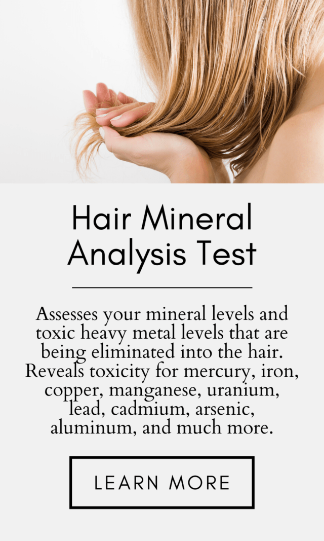
If you would like more information on what heavy metals and mineral deficiencies are present in your body and impeding your health, there’s a simple, affordable way to find out: hair mineral analysis (HTMA).
HTMA is a noninvasive lab test that measures both the heavy metal AND mineral content of your hair. Hair is used because it is where the body dumps heavy metals and excess minerals to protect the organs and maintain stable blood chemistry.
To get your own HTMA, discover your toxic metals levels, and mineral levels click here.
*Please note that A Hair Mineral Analysis (HTMA) is not intended to diagnose, treat, cure, reverse, or prevent any disease. It is not intended to replace any other medical test(s) that may be prescribed by your medical doctor. CitriCleanse is a dietary supplement that is not intended to diagnose, treat, cure, or prevent any disease. If you are dealing with infertility, please talk to your doctor before beginning any new supplement regimen.
Click Here for References+
- Mathur PP, D’Cruz SC. The effect of environmental contaminants on testicular function. Asian J Androl. 2011;13:585–591.
- Angelis, Cristina De, et al. “The Environment and Male Reproduction: The Effect of Cadmium Exposure on Reproductive Function and Its Implication in Fertility.” Reproductive Toxicology, vol. 73, 2017, pp. 105–127., doi:10.1016/j.reprotox.2017.07.021.
- Jameil NA. Maternal serum lead levels and risk of preeclampsia in pregnant women: a cohort study in a maternity hospital, Riyadh, Saudi Arabia. Int J Clin Exp Pathol. 2014;7:3182–3189.
- Motawei SM, Attalla SM, Gouda HE, El-Harouny MA, El-Mansoury AM. Lead level in pregnant women suffering from pre-eclampsia in Dakahlia, Egypt. Int J Occup Environ Med. 2013;4:36–44.
- Colquitt PJ. The effect of occupational exposure to mercury vapour on the fertility of female dental assistants. Occupational and Environmental Medicine. 1995;52:214.
- Cole DC, Wainman B, Sanin LH, Weber JP, Muggah H, Ibrahim S. Environmental contaminant levels and fecundability among non-smoking couples. Reproductive Toxicology. 2006;22:13–19.
- Burch JB, Wagner Robb S, Puett R, Cai B, Wilkerson R, Karmaus W, Vena J, Svendsen E. Mercury in fish and adverse reproductive outcomes: results from South Carolina. Int J Health Geogr. 2014;13:30.
- Ou L, Chen C, Chen L, Wang H, Yang T, Xie H, Tong Y, Hu D, Zhang W, Wang X. Low-level prenatal mercury exposure in north China: an exploratory study of anthropometric effects. Environ Sci Technol. 2015;49:6899–6908.
- Thomas S, Arbuckle TE, Fisher M, Fraser WD, Ettinger A, King W. Metals exposure and risk of small-for-gestational age birth in a Canadian birth cohort: The MIREC study. Environ Res. 2015;140:430–439.
- Chatterjee A, Chatterji U. Arsenic abrogates the estrogen-signaling pathway in the rat uterus. Reprod Biol Endocrinol. 2010;8:80.
- Chattopadhyay S, Ghosh D. The involvement of hypophyseal-gonadal and hypophyseal-adrenal axes in arsenic-mediated ovarian and uterine toxicity: modulation by hCG. J Biochem Mol Toxicol. 2010;24:29–41.
- Borja-Aburto VH, Hertz-Picciotto I, Rojas Lopez M, Farias P, Rios C, Blanco J. Blood lead levels measured prospectively and risk of spontaneous abortion. American Journal of Epidemiology. 1999;150:590–597.
- Quansah R, Armah FA, Essumang DK, Luginaah I, Clarke E, Marfoh K, Cobbina SJ, Nketiah-Amponsah E, Namujju PB, Obiri S, et al. Association of arsenic with adverse pregnancy outcomes/infant mortality: a systematic review and meta-analysis. Environ Health Perspect. 2015;123:412–421.
- Bloom MS, Surdu S, Neamtiu IA, Gurzau ES. Maternal arsenic exposure and birth outcomes: a comprehensive review of the epidemiologic literature focused on drinking water. Int J Hyg Environ Health. 2014;217:709–719.
- Huyck KL, Kile ML, Mahiuddin G, Quamruzzaman Q, Rahman M, Breton CV, Dobson CB, Frelich J, Hoffman E, Yousuf J, et al. Maternal arsenic exposure associated with low birth weight in Bangladesh. J Occup Environ Med. 2007;49:1097–1104.
- Laine JE, Bailey KA, Rubio-Andrade M, Olshan AF, Smeester L, Drobna Z, Herring AH, Styblo M, Garcia-Vargas GG, Fry RC. Maternal arsenic exposure, arsenic methylation efficiency, and birth outcomes in the Biomarkers of Exposure to Arsenic (BEAR) pregnancy cohort in Mexico. Environ Health Perspect. 2015;123:186–192.
- Laine JE, Bailey KA, Rubio-Andrade M, Olshan AF, Smeester L, Drobna Z, Herring AH, Styblo M, Garcia-Vargas GG, Fry RC. Maternal arsenic exposure, arsenic methylation efficiency, and birth outcomes in the Biomarkers of Exposure to Arsenic (BEAR) pregnancy cohort in Mexico. Environ Health Perspect. 2015;123:186–192.
- Ahmad SA, Sayed MH, Barua S, Khan MH, Faruquee MH, Jalil A, Hadi SA, Talukder HK. Arsenic in drinking water and pregnancy outcomes. Environmental Health Perspectives. 2001;109:629–631.
- Burch JB, Wagner Robb S, Puett R, Cai B, Wilkerson R, Karmaus W, Vena J, Svendsen E. Mercury in fish and adverse reproductive outcomes: results from South Carolina. Int J Health Geogr. 2014;13:30.
- Dallaire R, Dewailly E, Ayotte P, Forget-Dubois N, Jacobson SW, Jacobson JL, Muckle G. Exposure to organochlorines and mercury through fish and marine mammal consumption: associations with growth and duration of gestation among Inuit newborns. Environ Int. 2013;54:85–91.
- Ferguson KK, O’Neill MS, Meeker JD. Environmental contaminant exposures and preterm birth: a comprehensive review. J Toxicol Environ Health B Crit Rev. 2013;16:69–113.
- Perkins M, Wright RO, Amarasiriwardena CJ, Jayawardene I, Rifas-Shiman SL, Oken E. Very low maternal lead level in pregnancy and birth outcomes in an eastern Massachusetts population. Ann Epidemiol. 2014;24:915–919.
- Xue F, Holzman C, Rahbar MH, Trosko K, Fischer L. Maternal fish consumption, mercury levels, and risk of preterm delivery. Environmental Health Perspectives. 2007;115:42–47.
- Bloom MS, Surdu S, Neamtiu IA, Gurzau ES. Maternal arsenic exposure and birth outcomes: a comprehensive review of the epidemiologic literature focused on drinking water. Int J Hyg Environ Health. 2014;217:709–719.
- Myers SL, Lobdell DT, Liu Z, Xia Y, Ren H, Li Y, Kwok RK, Mumford JL, Mendola P. Maternal drinking water arsenic exposure and perinatal outcomes in inner Mongolia, China. Journal of Epidemiology and Community Health.
- Ferguson KK, O’Neill MS, Meeker JD. Environmental contaminant exposures and preterm birth: a comprehensive review. J Toxicol Environ Health B Crit Rev. 2013;16:69–113.
- Bermudez L, Garcia-Vicent C, Lopez J, Torro MI, Lurbe E. Assessment of ten trace elements in umbilical cord blood and maternal blood: association with birth weight. J Transl Med. 2015;13:291.
- Thomas S, Arbuckle TE, Fisher M, Fraser WD, Ettinger A, King W. Metals exposure and risk of small-for-gestational age birth in a Canadian birth cohort: The MIREC study. Environ Res. 2015;140:430–439.
- Bashore CJ, Geer LA, He X, Puett R, Parsons PJ, Palmer CD, Steuerwald AJ, Abulafia O, Dalloul M, Sapkota A. Maternal mercury exposure, season of conception and adverse birth outcomes in an urban immigrant community in Brooklyn, New York, U.S.A. Int J Environ Res Public Health. 2014;11:8414–8442.
- Laine JE, Bailey KA, Rubio-Andrade M, Olshan AF, Smeester L, Drobna Z, Herring AH, Styblo M, Garcia-Vargas GG, Fry RC. Maternal arsenic exposure, arsenic methylation efficiency, and birth outcomes in the Biomarkers of Exposure to ARsenic (BEAR) pregnancy cohort in Mexico. Environ Health Perspect. 2015;123:186–192.
- Ou L, Chen C, Chen L, Wang H, Yang T, Xie H, Tong Y, Hu D, Zhang W, Wang X. Low-level prenatal mercury exposure in north China: an exploratory study of anthropometric effects. Environ Sci Technol. 2015;49:6899–6908.
- Al-Saleh I, Shinwari N, Al-Amodi M. Accumulation of mercury in ovaries of mice after the application of skin-lightening creams. Biol Trace Elem Res. 2009;131:43–54.
- Al-Saleh I, Shinwari N, Mashhour A, Rabah A. Birth outcome measures and maternal exposure to heavy metals (lead, cadmium and mercury) in Saudi Arabian population. Int J Hyg Environ Health. 2014;217:205–218.
- Torres-Sanchez LE, Berkowitz G, Lopez-Carrillo L, Torres-Arreola L, Rios C, Lopez-Cervantes M. Intrauterine lead exposure and preterm birth. Environmental Research. 1999;81:297–301.
- Bloom MS, Surdu S, Neamtiu IA, Gurzau ES. Maternal arsenic exposure and birth outcomes: a comprehensive review of the epidemiologic literature focused on drinking water. Int J Hyg Environ Health. 2014;217:709–719.
- Sun, Jiantao, et al. “Heavy Metal Level in Human Semen with Different Fertility: a Meta-Analysis.” Biological Trace Element Research, vol. 176, no. 1, 2016, pp. 27–36., doi:10.1007/s12011-016-0804-2.
- Wang, Yi-Xin, et al. “Relationships between Seminal Plasma Metals/Metalloids and Semen Quality, Sperm Apoptosis and DNA Integrity.” Environmental Pollution, vol. 224, 2017, pp. 224–234., doi:10.1016/j.envpol.2017.01.083.
- Sun, Jiantao, et al. “Heavy Metal Level in Human Semen with Different Fertility: a Meta-Analysis.” Biological Trace Element Research, vol. 176, no. 1, 2016, pp. 27–36., doi:10.1007/s12011-016-0804-2.
- Cavallini A, Lippolis C, Vacca M, Nardelli C, Castegna A, Arnesano F, Carella N, Depalo R. The Effects of Chronic Lifelong Activation of the AHR Pathway by Industrial Chemical Pollutants on Female Human Reproduction. PLoS One. 2016;11:e0152181.
- Dickerson EH, Sathyapalan T, Knight R, Maguiness SM, Killick SR, Robinson J, Atkin SL. Endocrine disruptor & nutritional effects of heavy metals in ovarian hyperstimulation. J Assist Reprod Genet. 2011;28:1223–1228.
- Wright DL, Afeiche MC, Ehrlich S, Smith K, Williams PL, Chavarro JE, Batsis M, Toth TL, Hauser R. Hair mercury concentrations and in vitro fertilization (IVF) outcomes among women from a fertility clinic. Reprod Toxicol. 2015;51:125–132.
- Chattopadhyay S, Ghosh D. The involvement of hypophyseal-gonadal and hypophyseal-adrenal axes in arsenic-mediated ovarian and uterine toxicity: modulation by hCG. J Biochem Mol Toxicol. 2010;24:29–41.
- Al-Saleh I, Shinwari N, Al-Amodi M. Accumulation of mercury in ovaries of mice after the application of skin-lightening creams. Biol Trace Elem Res. 2009;131:43–54.
- Johnstone EB, Louis GM, Parsons PJ, Steuerwald AJ, Palmer CD, Chen Z, Sun L, Hammoud AO, Dorais J, Peterson CM. Increased urinary cobalt and whole blood concentrations of cadmium and lead in women with uterine leiomyomata: Findings from the ENDO Study. Reprod Toxicol. 2014;49:27–32.
- Chattopadhyay S, Ghosh D. The involvement of hypophyseal-gonadal and hypophyseal-adrenal axes in arsenic-mediated ovarian and uterine toxicity: modulation by hCG. J Biochem Mol Toxicol. 2010;24:29–41.
- Chatterjee A, Chatterji U. Arsenic abrogates the estrogen-signaling pathway in the rat uterus. Reprod Biol Endocrinol. 2010;8:80.
- Bloom MS, Parsons PJ, Steuerwald AJ, Schisterman EF, Browne RW, Kim K, et al. Toxic trace metals and human oocytes during in vitro fertilization (IVF). Reprod Toxicol 2010;29:298–305.
- Bloom MS, Fujimoto VY, Steuerwald AJ, Cheng G, Browne RW, Parsons Background exposure to toxic metals in women adversely influences pregnancy during in vitro fertilization (IVF). Reprod Toxicol 2012;34:471–81.
- Wang, Yi-Xin, et al. “Relationships between Seminal Plasma Metals/Metalloids and Semen Quality, Sperm Apoptosis and DNA Integrity.” Environmental Pollution, vol. 224, 2017, pp. 224–234., doi:10.1016/j.envpol.2017.01.083.
- Oliveira, H., Spano, M., Santos, C., Pereira Mde, L., 2009. Adverse effects of cadmium exposure on mouse sperm. Reprod. Toxicol. 28, 550e555.
- Pant, N., Kumar, R., Murthy, R.C., Srivastava, S.P., 2001. Male reproductive effect of arsenic in mice. Biometals 14, 113e117.
- Pant, N., Kumar, R., Murthy, R.C., Srivastava, S.P., 2001. Male reproductive effect of arsenic in mice. Biometals 14, 113e117.
- Wang, Y.X., Sun, , Feng, W., Wang, P., Yang, P., Li, J., Huang, Z., Chen, Y.J., Liu, C., Sun, L., Yue, J., Gu, L.J., Zeng, Q., Lu, W.Q., 2016b. Association of urinary metal levels with human semen quality: a cross-sectional study in China. Environ. Int. 91, 51e59.
- Meeker, J.D., Rossano, M.G., Protas, B., Diamond, P., Puscheck, E., Daly, D., Paneth, N., Wirth, J.J., 2008. Cadmium, lead, and other metals in relation to semen quality: human evidence for molybdenum as a male reproductive toxicant. Environ. Health Perspect. 116, 1473e1479.
- Benoff, , Hauser, R., Marmar, J.L., Hurley, I.R., Napolitano, B., Centola, G.M., 2009. Cadmium concentrations in blood and seminal plasma: correlations with sperm number and motility in three male populations (infertility patients, artificial insemination donors, and unselected volunteers). Mol. Med. 15, 248e262.
- Telisman, , Cvitkovic, P., Jurasovic, J., Pizent, A., Gavella, M., Rocic, B., 2000. Semen quality and reproductive endocrine function in relation to biomarkers of lead, cadmium, zinc, and copper in men. Environ. Health Perspect. 108, 45e53.
- Asadi, H., Zafari, F., Sarveazad, A., Abbasi, M., Safa, M., Koruji, M., Yari, A., Alizadeh Miran, R., 2014. Saffron improves epididymal sperm parameters in rats exposed to cadmium. Nephrourol. Mon. 6, e12125.
- Samikkannu, , Chen, C.H., Yih, L.H., Wang, A.S., Lin, S.Y., Chen, T.C., Jan, K.Y., 2003. Reactive oxygen species are involved in arsenic trioxide inhibition of pyruvate dehydrogenase activity. Chem. Res. Toxicol. 16, 409e414.
- Pant, N., Kumar, R., Murthy, R.C., Srivastava, S.P., 2001. Male reproductive effect of arsenic in mice. Biometals 14, 113e117.
- Zhu, X.H., Shen, Y.L., Jing, Y.K., Cai, X., Jia, P.M., Huang, Y., Tang, W., Shi, G.Y., Sun, Y.P., Dai, , Wang, Z.Y., Chen, S.J., Zhang, T.D., Waxman, S., Chen, Z., Chen, G.Q., 1999. Apoptosis and growth inhibition in malignant lymphocytes after treatment with arsenic trioxide at clinically achievable concentrations. J. Natl. Cancer Inst. 91, 772e778.
- Telisman, , Cvitkovic, P., Jurasovic, J., Pizent, A., Gavella, M., Rocic, B., 2000. Semen quality and reproductive endocrine function in relation to biomarkers of lead, cadmium, zinc, and copper in men. Environ. Health Perspect. 108, 45e53.
- Hernandez-Ochoa, I., Garcia-Vargas, G., Lopez-Carrillo, L., Rubio-Andrade, M., Moran-Martinez, J., Cebrian, M.E., Quintanilla-Vega, B., 2005. Low lead envi- ronmental exposure alters semen quality and sperm chromatin condensation in northern Mexico. Reprod. Toxicol. 20, 221e228.
- “National Primary Drinking Water Regulation Table.” EPA, Environmental Protection Agency, 21 Aug. 2017, https://www.epa.gov/ground-water-and-drinking-water/national-primary-drinking-water-regulation-table.
- Acosta, Izani Bonel, et al. “Effects of Exposure to Cadmium in Sperm Cells of Zebrafish, Danio Rerio.” Toxicology Reports, vol. 3, 2016, pp. 696–700., doi:10.1016/j.toxrep.2016.08.002.
- Olsson, Ing-Marie, et al. “Cadmium in Blood and Urine–Impact of Sex, Age, Dietary Intake, Iron Status, and Former Smoking–Association of Renal Effects.” Environmental Health Perspectives, vol. 110, no. 12, 2002, pp. 1185–1190., doi:10.1289/ehp.021101185.
- “Toxic Substances Portal – Cadmium.” Centers for Disease Control and Prevention, September 2012, https://www.atsdr.cdc.gov/toxprofiles/TP.asp?id=48&tid=15. Accessed 11/24/2019.
- Satarug, Soisungwan, and Michael R. Moore. “Adverse Health Effects of Chronic Exposure to Low-Level Cadmium in Foodstuffs and Cigarette Smoke.” Environmental Health Perspectives, vol. 112, no. 10, 2004, pp. 1099–1103., doi:10.1289/ehp.6751.
- Wier, Patrick J., et al. “Toxicity of Cadmium in the Perfused Human Placenta.” Toxicology and Applied Pharmacology, vol. 105, no. 1, 1990, pp. 156–171., doi:10.1016/0041-008x(90)90367-4.
- Piasek, Martina, et al. “Placental Cadmium and Progesterone Concentrations in Cigarette Smokers.” Reproductive Toxicology, vol. 15, no. 6, 2001, pp. 673–681., doi:10.1016/s0890-6238(01)00174-5.
- Staessen JA, Roels HA, Emelianov D, Kuznetsova T, Thijs L, Vangronsveld J, et Public Health and Environmental Exposure to Cadmium (PheeCad) Study Group. Environmental exposure to cadmium, forearm bone density, and risk of fractures: prospective population study. Lancet 1999;353: 1140–4.
- Bloom, Michael S., et al. “Toxic Trace Metals and Human Oocytes during in Vitro Fertilization (IVF).” Reproductive Toxicology, vol. 29, no. 3, 2010, pp. 298–305., doi:10.1016/j.reprotox.2010.01.003.
- Bloom, Michael S., et al. “Background Exposure to Toxic Metals in Women Adversely Influences Pregnancy during in Vitro Fertilization (IVF).” Reproductive Toxicology, vol. 34, no. 3, 2012, pp. 471–481., doi:10.1016/j.reprotox.2012.06.002.
- Gardner RM, Kippler M, Tofail F, Bottai M, Hamadani J, Grand‘er M, et Environmental exposure to metals and children’s growth to age 5 years: a prospective cohort study. Am J Epidemiol 2013;177:1356–67.
- Segal, Thalia R., and Linda C. Giudice. “Before the Beginning: Environmental Exposures and Reproductive and Obstetrical Outcomes.” Fertility and Sterility, vol. 112, no. 4, 2019, pp. 613–621., doi:10.1016/j.fertnstert.2019.08.001.
- Kippler, Maria, et al. “Maternal Cadmium Exposure during Pregnancy and Size at Birth: A Prospective Cohort Study.” Environmental Health Perspectives, vol. 120, no. 2, 2012, pp. 284–289., doi:10.1289/ehp.1103711.
- Kippler, Maria, et al. “Environmental Exposure to Arsenic and Cadmium during Pregnancy and Fetal Size: A Longitudinal Study in Rural Bangladesh.” Reproductive Toxicology, vol. 34, no. 4, 2012, pp. 504–511., doi:10.1016/j.reprotox.2012.08.002.
- Lin, C.-M., et al. “Does Prenatal Cadmium Exposure Affect Fetal and Child Growth?” Occupational and Environmental Medicine, vol. 68, no. 9, 2010, pp. 641–646., doi:10.1136/oem.2010.059758.
- Salpietro, C. D., et al. “Cadmium Concentration in Maternal and Cord Blood and Infant Birth Weight: a Study on Healthy Non-Smoking Women.” Journal of Perinatal Medicine, vol. 30, no. 5, 2002, doi:10.1515/jpm.2002.061.
- Gardner RM, Kippler M, Tofail F, Bottai M, Hamadani J, Grand‘er M, et Environmental exposure to metals and children’s growth to age 5 years: a prospective cohort study. Am J Epidemiol 2013;177:1356–67.
- Staessen JA, Roels HA, Emelianov D, Kuznetsova T, Thijs L, Vangronsveld J, et Public Health and Environmental Exposure to Cadmium (PheeCad) Study Group. Environmental exposure to cadmium, forearm bone density, and risk of fractures: prospective population study. Lancet 1999;353: 1140–4.
- Kippler, Maria, et al. “Sex-Specific Effects of Early Life Cadmium Exposure on DNA Methylation and Implications for Birth Weight.” Epigenetics, vol. 8, no. 5, 2013, pp. 494–503., doi:10.4161/epi.24401.
- Vilahur N, Vahter M, Broberg The epigenetic effects of prenatal cad-mium exposure. Curr Environ Health Rep 2015;2:195–203.
- Boeke CE, Baccarelli A, Kleinman KP, Burris HH, Litonjua AA, Rifas- Shiman SL, et Gestational intake of methyl donors and global LINE-1 DNA methylation in maternal and cord blood: prospective results from a folate-replete population. Epigenetics 2012;7:253–60.
- Sanders, Alison, et al. “Cadmium Exposure and the Epigenome: Exposure-Associated Patterns of DNA Methylation in Leukocytes from Mother-Baby Pairs.” Epigenetics, vol. 9, no. 2, 2013, pp. 212–221., doi:10.4161/epi.26798.
- Pizent, Alica, et al. “Reproductive Toxicity of Metals in Men.” Archives of Industrial Hygiene and Toxicology, vol. 63, no. Supplement-1, Jan. 2012, doi:10.2478/10004-1254-63-2012-2151.
- Kumar S, Sharma Cadmium toxicity: effects on human reproduction and fertility. Rev Environ Health 2019; https://doi.org/10.1515/reveh-2019-0016.
- Monsefi, Malihezaman, et al. “Cadmium-Induced Infertility in Male Mice.” Environmental Toxicology, 2009, doi:10.1002/tox.20468.
- Acosta, Izani Bonel, et al. “Effects of Exposure to Cadmium in Sperm Cells of Zebrafish, Danio Rerio.” Toxicology Reports, vol. 3, 2016, pp. 696–700., doi:10.1016/j.toxrep.2016.08.002.
- Babaknejad, Nasim, et al. “Cadmium Testicular Toxicity in Male Wistar Rats: Protective Roles of Zinc and Magnesium.” Biological Trace Element Research, vol. 185, no. 1, 2017, pp. 106–115., doi:10.1007/s12011-017-1218-5.
- Marchiani, S, et al. “Acute Effects on Human Sperm Exposed in Vitro to Cadmium Chloride and Diisobutyl Phthalate.” Reproduction, vol. 158, no. 3, 2019, pp. 281–290., doi:10.1530/rep-19-0207.
- Sukhn, Carol, et al. “Associations of Semen Quality with Non-Essential Heavy Metals in Blood and Seminal Fluid: Data from the Environment and Male Infertility (EMI) Study in Lebanon.” Journal of Assisted Reproduction and Genetics, vol. 35, no. 9, 2018, pp. 1691–1701., doi:10.1007/s10815-018-1236-z.
- Angelis, Cristina De, et al. “The Environment and Male Reproduction: The Effect of Cadmium Exposure on Reproductive Function and Its Implication in Fertility.” Reproductive Toxicology, vol. 73, 2017, pp. 105–127., doi:10.1016/j.reprotox.2017.07.021.
- Babaknejad, Nasim, et al. “Cadmium Testicular Toxicity in Male Wistar Rats: Protective Roles of Zinc and Magnesium.” Biological Trace Element Research, vol. 185, no. 1, 2017, pp. 106–115., doi:10.1007/s12011-017-1218-5.
- Benoff, Susan, et al. “Cadmium Concentrations in Blood and Seminal Plasma: Correlations with Sperm Number and Motility in Three Male Populations (Infertility Patients, Artificial Insemination Donors, and Unselected Volunteers).” Molecular Medicine, vol. 15, no. 7-8, 2009, pp. 248–262., doi:10.2119/molmed.2008.00104.
- Mendiola, Jaime, et al. “Relationships between Heavy Metal Concentrations in Three Different Body Fluids and Male Reproductive Parameters: a Pilot Study.” Environmental Health, vol. 10, no. 1, 2011, doi:10.1186/1476-069x-10-6.
- Pant, N., et al. “Correlation between Lead and Cadmium Concentration and Semen Quality.” Andrologia, 2014, doi:10.1111/and.12342.
- Dawson, Earl B., et al. “Comparison of Sperm Viability with Seminal Plasma Metal Levels.” Biological Trace Element Research, vol. 64, no. 1-3, 1998, pp. 215–219., doi:10.1007/bf02783337.
- Xu, LC, et al. “Effects of cadmium on rat sperm motility evaluated with computer assisted sperm analysis.” Biomedical Environmental Sciences, vol. 14, no. 3, 2001, pp. 312–317.
- Hew, K.w., et al. “A Single Low Cadmium Dose Causes Failure of Spermiation in the Rat.” Toxicology and Applied Pharmacology, vol. 121, no. 1, 1993, pp. 15–21., doi:10.1006/taap.1993.1123.
- El-Demerdash, Fatma M, et al. “Cadmium-Induced Changes in Lipid Peroxidation, Blood Hematology, Biochemical Parameters and Semen Quality of Male Rats: Protective Role of Vitamin E and β-Carotene.” Food and Chemical Toxicology, vol. 42, no. 10, 2004, pp. 1563–1571., doi:10.1016/j.fct.2004.05.001.
- Oliveira, Helena, et al. “Adverse Effects of Cadmium Exposure on Mouse Sperm.” Reproductive Toxicology, vol. 28, no. 4, 2009, pp. 550–555., doi:10.1016/j.reprotox.2009.08.001.
- Sierra-Marquez, Lucellys, et al. “Effects of Cadmium Exposure on Sperm and Larvae of the Neotropical Fish Prochilodus Magdalenae.” Comparative Biochemistry and Physiology Part C: Toxicology & Pharmacology, vol. 225, 2019, p. 108577., doi:10.1016/j.cbpc.2019.108577.
- Abarikwu, S. O., et al. “Rutin, an Antioxidant Flavonoid, Induces Glutathione and Glutathione Peroxidase Activities to Protect against Ethanol Effects in Cadmium-Induced Oxidative Stress in the Testis of Adult Rats.” Andrologia, vol. 49, no. 7, 2016, doi:10.1111/and.12696.
- Akinloye, O, et al. “Cadmium toxicity: a possible cause of male infertility in Nigeria.” Reproductive Biolology, vol. 6, 2006, pp. 17-30.
- Yang, Hong, and Yan Shu. “Cadmium Transporters in the Kidney and Cadmium-Induced Nephrotoxicity.” International Journal of Molecular Sciences, vol. 16, no. 1, Sept. 2015, pp. 1484–1494., doi:10.3390/ijms16011484.
- Minutoli, Letteria, et al. “Flavocoxid Protects Against Cadmium-Induced Disruption of the Blood–Testis Barrier and Improves Testicular Damage and Germ Cell Impairment in Mice.” Toxicological Sciences, vol. 149, no. 1, Mar. 2015, pp. 270–270., doi:10.1093/toxsci/kfv247.
- Zhao, Li-Lin, et al. “Reproductive Effects of Cadmium on Sperm Function and Early Embryonic Development in Vitro.” Plos One, vol. 12, no. 11, Feb. 2017, doi:10.1371/journal.pone.0186727.
- Martino, N.a., et al. “Exposure to Cadmium during in Vitro Maturation at Environmental Nanomolar Levels Impairs Oocyte Fertilization through Oxidative Damage: A Large Animal Model Study.” Reproductive Toxicology, vol. 69, 2017, pp. 132–145., doi:10.1016/j.reprotox.2017.02.005.
- Cheng, Yuyao, et al. “Reproductive Toxicity of Acute Cd Exposure in Mouse: Resulting in Oocyte Defects and Decreased Female Fertility.” Toxicology and Applied Pharmacology, vol. 379, 2019, p. 114684., doi:10.1016/j.taap.2019.114684.
- Vine, M. F. “Smoking and Male Reproduction: a Review.” International Journal of Andrology, vol. 19, no. 6, 1996, pp. 323–337., doi:10.1111/j.1365-2605.1996.tb00523.x.
- Bloom, Michael S., et al. “Toxic Trace Metals and Human Oocytes during in Vitro Fertilization (IVF).” Reproductive Toxicology, vol. 29, no. 3, 2010, pp. 298–305., doi:10.1016/j.reprotox.2010.01.003.
- Bloom, Michael S., et al. “Background Exposure to Toxic Metals in Women Adversely Influences Pregnancy during in Vitro Fertilization (IVF).” Reproductive Toxicology, vol. 34, no. 3, 2012, pp. 471–481., doi:10.1016/j.reprotox.2012.06.002.
- Zhao, Li-Lin, et al. “Reproductive Effects of Cadmium on Sperm Function and Early Embryonic Development in Vitro.” Plos One, vol. 12, no. 11, Feb. 2017, doi:10.1371/journal.pone.0186727.
- Zhu, Jia-Qiao, et al. “Cadmium Exposure of Female Mice Impairs the Meiotic Maturation of Oocytes and Subsequent Embryonic Development.” Toxicological Sciences, vol. 164, no. 1, 2018, pp. 289–299., doi:10.1093/toxsci/kfy089.
- Zhu, Jia-Qiao, et al. “Cadmium Exposure of Female Mice Impairs the Meiotic Maturation of Oocytes and Subsequent Embryonic Development.” Toxicological Sciences, vol. 164, no. 1, 2018, pp. 289–299., doi:10.1093/toxsci/kfy089.
- Zhu, Jia-Qiao, et al. “Cadmium Exposure of Female Mice Impairs the Meiotic Maturation of Oocytes and Subsequent Embryonic Development.” Toxicological Sciences, vol. 164, no. 1, 2018, pp. 289–299., doi:10.1093/toxsci/kfy089.
- Paksy, Katalin, et al. “Acute Effects of Cadmium on Preovulatory Serum FSH, LH, and Prolactin Levels and on Ovulation and Ovarian Hormone Secretion in Estrous Rats.” Reproductive Toxicology, vol. 3, no. 4, 1989, pp. 241–247., doi:10.1016/0890-6238(89)90018-x.
- Piasek, Martina, and John W. Laskey. “Acute Cadmium Exposure and Ovarian Steroidogenesis in Cycling and Pregnant Rats.” Reproductive Toxicology, vol. 8, no. 6, 1994, pp. 495–507., doi:10.1016/0890-6238(94)90032-9.
- Thompson, J, and J Bannigan. “Cadmium: Toxic Effects on the Reproductive System and the Embryo.” Reproductive Toxicology, vol. 25, no. 3, 2008, pp. 304–315., doi:10.1016/j.reprotox.2008.02.001.
- “Toxic Substances Portal – Arsenic.” Centers for Disease Control and Prevention, , August 2007, https://www.atsdr.cdc.gov/toxprofiles/tp.asp?id=22&tid=3. Accessed 11/24/2019.
- Zhu, Jia-Qiao, et al. “Cadmium Exposure of Female Mice Impairs the Meiotic Maturation of Oocytes and Subsequent Embryonic Development.” Toxicological Sciences, vol. 164, no. 1, 2018, pp. 289–299., doi:10.1093/toxsci/kfy089.
- Ferguson KK, O’Neill MS, Meeker JD. Environmental contaminant exposures and preterm birth: a comprehensive review. J Toxicol Environ Health B Crit Rev. 2013; 16:69–113.
- García-Fortea, Pedro, et al. “Toxic Elements in Hair and in Vitro Fertilization Outcomes: A Prospective Cohort Study.” Reproductive Toxicology, vol. 77, 2018, pp. 43–52., doi:10.1016/j.reprotox.2018.02.001.
- Li, Ping, et al. “Seminal plasma metals concentration with respect to semen quality.” Biological Trace Elements Research, vol 148, no. 1, July 2012, pp. 1-6., doi: 10.1007/s12011-012-9335-7.
- J Physiol Pharmacol.2019 Jun;70(3). doi: 10.26402/jpp.2019.3.02. Epub 2019 Sep 18.
- Toxic Substances Portal – Arsenic.” Centers for Disease Control and Prevention, , August 2007, https://www.atsdr.cdc.gov/toxprofiles/tp.asp?id=22&tid=3. Accessed 11/24/2019.
- Hamada, Alaa, Esteves, Sandro C., Nizza, Mark, & Agarwal, Ashok. (2012). Unexplained Male infertility: diagnosis and Management. International braz j urol, 38(5), 576-594. https://dx.doi.org/10.1590/S1677-55382012000500002
- Arsenic Metabolites in Human Urine after Ingestion of an ArsenosugaKevin A. Francesconi, René Tanggaar, Christine J. McKenzie, Walter Goessler Clinical Chemistry Jan 2002, 48 (1) 92-101.
- Valenzuela OL, Borja-Aburto VH, Garcia-Vargas GG, Cruz-Gonzalez MB, Garcia-Montalvo EA, Calderon-Aranda ES, Del Razo LM. Urinary trivalent methylated arsenic species in a population chronically exposed to inorganic arsenic. Environ Health Perspect. 2005 Mar;113(3):250-4.
- Shen, H., Xu,W., Zhang, J., Chen, M., Martin, F.L., Xia, Y., et al., 2013. Urinary metabolic biomarkers link oxidative stress indicators associated with general arsenic exposure to male infertility in a Han Chinese population. Environ. Sci. Technol. 47, 8843–8851.
- Wang, Chao, et al. “The Classic EDCs, Phthalate Esters and Organochlorines, in Relation to Abnormal Sperm Quality: a Systematic Review with Meta-Analysis.” Scientific Reports, vol. 6, no. 1, 2016, doi:10.1038/srep19982.
- “Toxic Substances Portal – Mercury.” Centers for Disease Control and Prevention, Centers for Disease Control and Prevention, https://www.atsdr.cdc.gov/toxfaqs/TF.asp?id=113&tid=24#bo
- “Toxic Substances Portal – Mercury.” Centers for Disease Control and Prevention, Centers for Disease Control and Prevention, https://www.atsdr.cdc.gov/toxfaqs/TF.asp?id=113&tid=24#bo
- Ferguson KK, O’Neill MS, Meeker JD. Environmental contaminant exposures and preterm birth: a comprehensive review. J Toxicol Environ Health B Crit Rev. 2013;16:69–113.
- “Toxic Substances Portal – Mercury.” Centers for Disease Control and Prevention, Centers for Disease Control and Prevention, https://www.atsdr.cdc.gov/toxfaqs/TF.asp?id=113&tid=24#bo
- “Toxic Substances Portal – Mercury.” Centers for Disease Control and Prevention, Centers for Disease Control and Prevention, https://www.atsdr.cdc.gov/toxfaqs/TF.asp?id=113&tid=24#bo
- “Health Effects of Exposures to Mercury.” EPA, Environmental Protection Agency, 10 May 2019, https://www.epa.gov/mercury/health-effects-exposures-mercury.
- Maeda, Eri, et al. “Associations of Environmental Exposures to Methylmercury and Selenium with Female Infertility: A Case–Control Study.” Environmental Research, vol. 168, 2019, pp. 357–363., doi:10.1016/j.envres.2018.10.007.
- García-Fortea, Pedro, et al. “Toxic Elements in Hair and in Vitro Fertilization Outcomes: A Prospective Cohort Study.” Reproductive Toxicology, vol. 77, 2018, pp. 43–52., doi:10.1016/j.reprotox.2018.02.001.
- Dickerson EH, Sathyapalan T, Knight R, Maguiness SM, Killick SR, Robinson J, Atkin SL. Endocrine disruptor & nutritional effects of heavy metals in ovarian hyperstimulation. J Assist Reprod 2011; 28:1223–1228. [PubMed: 22071884]
- Maeda, Eri, et al. “Associations of Environmental Exposures to Methylmercury and Selenium with Female Infertility: A Case–Control Study.” Environmental Research, vol. 168, 2019, pp. 357–363., doi:10.1016/j.envres.2018.10.007.
- Wright DL, Afeiche MC, Ehrlich S, Smith K, Williams PL, Chavarro JE, Batsis M, Toth TL, Hauser R. Hair mercury concentrations and in vitro fertilization (IVF) outcomes among women from a fertility clinic. Reprod Toxicol. 2015; 51:125–132. [PubMed: 25601638]
- Bloom MS, Surdu S, Neamtiu IA, Gurzau ES. Maternal arsenic exposure and birth outcomes: a comprehensive review of the epidemiologic literature focused on drinking water. Int J Hyg Environ Health. 2014;217:709–719.
- Xue F, Holzman C, Rahbar MH, Trosko K, Fischer L. Maternal fish consumption, mercury levels, and risk of preterm delivery. Environmental Health Perspectives. 2007;115:42–47.
- Ahmad SA, Sayed MH, Barua S, Khan MH, Faruquee MH, Jalil A, Hadi SA, Talukder HK. Arsenic in drinking water and pregnancy outcomes. Environmental Health Perspectives. 2001; 109:629–631. [PubMed: 11445518]
- Burch JB, Wagner Robb S, Puett R, Cai B, Wilkerson R, Karmaus W, Vena J, Svendsen E. Mercury in fish and adverse reproductive outcomes: results from South Carolina. Int J Health Geogr. 2014; 13:30. [PubMed: 25127892]
- Dallaire R, Dewailly E, Ayotte P, Forget-Dubois N, Jacobson SW, Jacobson JL, Muckle G. Exposure to organochlorines and mercury through fish and marine mammal consumption: associations with growth and duration of gestation among Inuit newborns. Environ Int. 2013; 54:85–91. [PubMed: 23422685]
- Perkins M, Wright RO, Amarasiriwardena CJ, Jayawardene I, Rifas-Shiman SL, Oken E. Very low maternal lead level in pregnancy and birth outcomes in an eastern Massachusetts population. Ann Epidemiol. 2014; 24:915–919. [PubMed: 25444892]
- “Toxic Substances Portal – Lead.” Centers for Disease Control and Prevention, May 2019, https://www.atsdr.cdc.gov/substances/toxsubstance.asp?toxid=22. Accessed 11/24/2019.
- “Toxic Substances Portal – Lead.” Centers for Disease Control and Prevention, May 2019, https://www.atsdr.cdc.gov/substances/toxsubstance.asp?toxid=22. Accessed 11/24/2019.
- Ferguson KK, O’Neill MS, Meeker JD. Environmental contaminant exposures and preterm birth: a comprehensive review. J Toxicol Environ Health B Crit Rev. 2013;16:69–113.
- Spanier AJ, Wilson S, Ho M, Hornung R, Lanphear The contribution of housing renovation to children’s blood lead levels: a cohort study. Environ Health 2013;12:72. https://ehjournal.biomedcentral.com/articles/10.1186/1476-069X-12-72
- “Toxic Substances Portal – Lead.” Centers for Disease Control and Prevention, May 2019, https://www.atsdr.cdc.gov/substances/toxsubstance.asp?toxid=22. Accessed 11/24/2019.
- “Toxic Substances Portal – Lead.” Centers for Disease Control and Prevention, May 2019, https://www.atsdr.cdc.gov/substances/toxsubstance.asp?toxid=22. Accessed 11/24/2019.
- “Toxic Substances Portal – Lead.” Centers for Disease Control and Prevention, May 2019, https://www.atsdr.cdc.gov/substances/toxsubstance.asp?toxid=22. Accessed 11/24/2019.
- Louis, G. M. Buck, et al. “Paternal Exposures to Environmental Chemicals and Time-to-Pregnancy: Overview of Results from the LIFE Study.” Andrology, vol. 4, no. 4, July 2016, pp. 639–647., doi:10.1111/andr.12171.
- Li, Ping & Zhong, Yuanfu & Jiang, Xiaoming & Wang, Chonggang & Zuo, Zhenghong & Sha, Aiguo. (2012). Seminal Plasma Metals Concentration with Respect to Semen Quality. Biological trace element research. 148. 1-6. 10.1007/s12011-012-9335-7.
- Kumar, Sunil. “Occupational and Environmental Exposure to Lead and Reproductive Health Impairment: An Overview.” Indian Journal of Occupational and Environmental Medicine, vol 22, no. 3, 2018, pp 128-137., doi: 10.4103/ijoem.IJOEM_126_18: 10.4103/ijoem.IJOEM_126_18.
- Marzec-Wróblewska, Urszula, et al. “Human Sperm Characteristics with Regard to Cobalt, Chromium, and Lead in Semen and Activity of Catalase in Seminal Plasma.” Biological Trace Element Research, vol. 188, no. 2, 2018, pp. 251–260., doi:10.1007/s12011-018-1416-9.
- Kim, Keewan, et al. “Toxic Metals in Seminal Plasma and in vitro Fertilization (IVF) Outcomes.” Environmental Research, vol. 133, pp. 334-337. Doi: 10.1016/j.envres.2014.06.014.
- Li, Cuiling, et al. “Lead Exposure Reduces Sperm Quality and DNA Integrity in Mice.” Environmental Toxicology, vol. 33, no. 5, 2018, pp. 594–602., doi:10.1002/tox.22545.
- “Toxic Substances Portal – Aluminum.” Centers for Disease Control and Prevention, September 2008. https://www.atsdr.cdc.gov/toxprofiles/tp.asp?id=191&tid=34. Accessed 11/25/2019.
- “Toxic Substances Portal – Aluminum.” Centers for Disease Control and Prevention, September 2008. https://www.atsdr.cdc.gov/toxprofiles/tp.asp?id=191&tid=34. Accessed 11/25/2019.
- Martinez, Caroline Silvera, et al. “Aluminum Exposure for 60 days at Human Dietary Levels Impairs Spermatogenesis and Sperm Quality in Rats.” Reproductive Toxicology, vol. 73, pp. 128-141.
- Klein, J.P., et al. “Aluminum Content of Human Semen: Implications for Semen Quality.” Reproductive Toxicology, vol 50, pp. 43-48.
- Sun, Jiantao, et al. “Heavy Metal Level in Human Semen with Different Fertility: a Meta-Analysis.” Biological Trace Element Research, vol. 176, no. 1, 2016, pp. 27–36., doi:10.1007/s12011-016-0804-2.
- Squibb KS, McDiarmid MA. Depleted uranium exposure and health effects in Gulf War Philosophical transactions of the Royal Society of London Series B Biol Sci. 2006;361(1468): 639–48.
- Sukhn, Carol, et al. “Associations of Semen Quality with Non-Essential Heavy Metals in Blood and Seminal Fluid: Data from the Environment and Male Infertility (EMI) Study in Lebanon.” Journal of Assisted Reproduction and Genetics, vol. 35, no. 9, 2018, pp. 1691–1701., doi:10.1007/s10815-018-1236-z.
- Llobet JM, Sirvent JJ, Ortega A, Domingo Influence of chronic exposure to uranium on male reproduction in mice. Fundam Appl Toxicol. 1991;16(4):821–9.
- National Research Council (US) Committee on Diet and Health. “Diet and Health: Implications for Reducing Chronic Disease Risk.” Washington (DC): National Academies Press (US); 1989.
- National Research Council (US) Committee on Diet and Health. “Diet and Health: Implications for Reducing Chronic Disease Risk.” Washington (DC): National Academies Press (US); 1989.
- Li, Ping & Zhong, Yuanfu & Jiang, Xiaoming & Wang, Chonggang & Zuo, Zhenghong & Sha, Aiguo. (2012). Seminal Plasma Metals Concentration with Respect to Semen Quality. Biological trace element research. 148. 1-6. 10.1007/s12011-012-9335-7.
- Bergamo, Paolo, et al. “Human Semen as an Early, Sensitive Biomarker of Highly Polluted Living Environment in Healthy Men: A Pilot Biomonitoring Study on Trace Elements in Blood and Semen and Their Relationship with Sperm Quality and RedOx Status.” Reproductive Toxicology, vol. 66, 2016, pp. 1–9., doi:10.1016/j.reprotox.2016.07.018.
- Dobrakowski, Michał, et al. “The Role of Oxidative Stress, Selected Metals, and Parameters of the Immune System in Male Fertility.” Oxidative Medicine and Cellular Longevity, vol. 2018, Apr. 2018, pp. 1–8., doi:10.1155/2018/6249536.
- Kasperczyk, Aleksandra & Dobrakowski, Michał & Horak, Stanisław & Zalejska-Fiolka, Jolanta & Birkner, Ewa. (2014). The influence of macro and trace elements on sperm quality. Journal of trace elements in medicine and biology : organ of the Society for Minerals and Trace Elements (GMS). 30. 10.1016/j.jtemb.2014.12.007.
- “Toxic Substances Portal – Chromium.” Centers for Disease Control and Prevention, March 2011. https://www.atsdr.cdc.gov/substances/toxsubstance.asp?toxid=17
- Marzec-Wróblewska, Urszula, et al. “Human Sperm Characteristics with Regard to Cobalt, Chromium, and Lead in Semen and Activity of Catalase in Seminal Plasma.” Biological Trace Element Research, vol. 188, no. 2, 2018, pp. 251–260., doi:10.1007/s12011-018-1416-9.
- Ma, Yanmin, et al. “Effects of Environmental Contaminants on Fertility and Reproductive Health.” Journal of Environmental Sciences, vol. 77, 2019, pp. 210–217., doi:10.1016/j.jes.2018.07.015.
- Marzec-Wróblewska, Urszula, et al. “Human Sperm Characteristics with Regard to Cobalt, Chromium, and Lead in Semen and Activity of Catalase in Seminal Plasma.” Biological Trace Element Research, vol. 188, no. 2, 2018, pp. 251–260., doi:10.1007/s12011-018-1416-9.