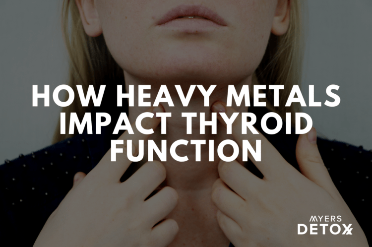
How Heavy Metals Impact Thyroid Function
Hypothyroidism is low thyroid function. It is one of the most common chronic conditions affecting Americans today, with an estimated 20 million people diagnosed with the disease. Millions more are undiagnosed.
While every cell in the body depends on thyroid hormones to function, this butterfly-shaped gland in your throat area is also very vulnerable to heavy metals and environmental toxins. As a result, heavy metals and toxins could sap your energy and well being by damaging thyroid function. Read on to learn more about how heavy metals impact thyroid function.
What you’ll learn in this article:
- The thyroid gland is very vulnerable to damage from heavy metals and environmental toxins.
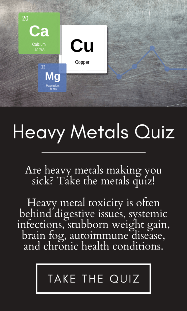
- The heavy metals that have been shown to have the largest impact on the thyroid are cadmium, lead, mercury, and aluminum.
- Halogens such as bromine in fire retardants and other environmental toxins have been shown to impact thyroid function.
- Every cell in the body needs thyroid hormone to function.
- Hypothyroidism causes fatigue, mood issues, weight gain, hair loss, and other symptoms.
- Ninety percent of hypothyroidism cases are caused by an autoimmune disease called Hashimoto’s.
- You can safely and gently detox your thyroid tissue from heavy metals and environmental toxins with pure, natural compounds and follow the program daily as a preventive measure.
Can’t Lose Weight? See If You Have These Other Hypothyroidism Symptoms
Hypothyroidism means the thyroid gland is underactive. It operates like the gas pedal for your metabolism, giving cells the energy they need to function.
This includes cells that keep your fat burning, your brain functioning, your gut moving things along, and your body’s hormones, organs and systems operating.
It’s no wonder people with hypothyroidism feel so terrible all the time. See if these common hypothyroidism symptoms (1) apply to you:
- Fatigue
- Low motivation
- Need to sleep excessively
- Hair loss and thinning
- Muscle weakness
- Feel cold all the time
- Constipation
- Dry skin
- Weight gain
- Inability to lose weight, even with low calorie dieting and exercise
- Brain fog and memory loss
- Puffy face and eyes
- Hoarse voice
- High cholesterol
- Muscle aches, tenderness, and stiffness
- Stiff and painful joints
Doctors typically diagnose hypothyroidism when they see a high level of thyroid stimulating hormone (TSH) on a blood test and prescribe thyroid hormone medication.
More astute doctors and patients know that 90 percent of hypothyroidism cases in the US are caused by Hashimoto’s, and autoimmune disease that attacks and damages the thyroid gland.
Why You Need to Detox Your Thyroid of Heavy Metals and Toxins
Although treating hypothyroidism requires a multi-faceted approach that frequently involves medication, one factor typically overlooked is the role heavy metals and toxins play in the health and function of the thyroid gland.
In fact, multiple studies demonstrate the impact of these compounds on thyroid function, which I discuss below.
Although thyroid medication may be necessary, managing your hypothyroidism typically also requires adapting healthy lifestyle and dietary habits, as well as undergoing a thyroid detox.
Check out these alarming studies on the effects of heavy metals and toxins on our delicate but powerful thyroid gland. When I became aware I had thyroid issues I sought out the underlying root cause of why I had low thyroid function (toxins) in an effort to restore function and hormone production.
How Heavy Metals Impact Thyroid Function
First, let’s look at how heavy metals and toxins in general impact the thyroid. The heavy metals that have been shown to have the largest impact on the thyroid are cadmium, lead, mercury, and aluminum.
Toxic heavy metals (2) circulating in the bloodstream can make their way into thyroid tissue and reduce thyroid function in a variety of ways:
They inhibit enzyme function. Enzymes are catalysts, or spark plugs, throughout the body that are necessary to synthesize or convert thyroid hormones and other compounds the body needs to operate. The interference of heavy metals in enzymes function has been shown to hinder the production of thyroglobulin, a storage form of thyroid hormone. Metals also interfere in the conversion of T4 to T3.
They interfere with the absorption of amino acids necessary for the production of thyroid hormones. Toxic metals such as arsenic, aluminum, tin, and thallium have been shown to hinder the absorption of tyrosine. Tyrosine is an amino acid — or building block protein — that is necessary for the synthesis of thyroid hormones. Additionally, proton pump inhibitor medications taken for acid reflux or GERD can especially deplete your thyroid of tyrosine (3).
They sabotage cellular energy necessary for thyroid hormone production. Toxic metals such as arsenic, aluminum, tin and thallium interfere with energy production inside the body’s cells. Though it’s a small gland, the thyroid has a big job of supplying the body’s cells with energy via thyroid hormone. The uptake of iodine by the gland and the production of thyroid hormones are very demanding of energy. When toxic heavy metals interfere with cellular energy, this in turn can hinder the production of thyroid hormones.
How Mercury Impacts Thyroid Function
Mercury contaminates the environment through coal burning, waste incineration, and other industrial and manufacturing processes. Dietary contamination through consumption of large fish such as tuna, swordfish, shark, tilefish, mackerel, marlin, orange roughy, and cod is another common source of exposure.
Mercury is also found in products such as topical skin lightening creams sold outside the United States. Let’s not forget that many get daily exposure from past and present mercury amalgam fillings.
Mercury is associated with lowering thyroid hormone levels by an indirect effect on the hypothalamus, the area of the brain responsible for coordinating hormone function and which can collect mercury deposits that make their way into the brain.
The hypothalamus stays in constant contact with the thyroid gland and the rest of the body to regulate the production and release of thyroid hormone. It does this by releasing a thyroid signaling hormone called thyrotropin releasing hormone (TRH).
When mercury deposits bog down cellular function in the hypothalamus, its ability to communicate with the thyroid gland suffers (4). As a result, the thyroid is not activated to produce enough thyroid hormone and overall thyroid activity lowers (5).
Additionally, mercury deposits itself in the pituitary gland, a pea-sized gland at the bottom of the brain that controls the function of the body’s hormonal glands. These mercury deposits prevent the synthesis of TSH by the pituitary, the hormone necessary to tell the thyroid to make thyroid hormone the cells can use.
The thyroid gland makes primarily T4, a thyroid hormone that is inactive and must be converted to T3, an active form of thyroid hormone the body’s cells can use. This conversion takes place throughout the body but predominantly in the liver and the gut.
Mercury, however, interferes in the conversion of T4 to T3 by poisoning the enzymes necessary for this conversion (6). This depletes cells of T3 and symptoms of hypothyroidism ensue.
Lastly, because mercury is structurally similar to iodine, it competes with iodine in the thyroid gland (7). As a result, the thyroid gland can become mercury toxic and prevent the production of T4 and T3, leading to hypothyroidism symptoms.
Mercury and Autoimmune Hashimoto’s
As I mentioned earlier, 90 percent of hypothyroid cases are caused by an autoimmune disease called Hashimoto’s. One study showed that women with elevated levels of mercury were twice as likely to test positive for antibodies to the thyroid, a marker that indicates the development of Hashimoto’s (8).
Mercury has been associated in the research with triggering autoimmune diseases such as Hashimoto’s. A 2015 study found that even low levels of mercury generally considered safe were associated with a higher rate of “autoantibodies,” or antibodies to tissue in the body (9). This is a marker for either autoimmune disease or an autoimmune process that could lead to disease.
Although the reasons mercury can trigger autoimmunity are not fully understood, we do know that the immune system can develop a sensitivity to mercury that has bonded with tissues (10). The immune system does not recognize the mercury-laden tissue and responds to it as a foreign invader. This inadvertently triggers an immune response both to the tissue and the mercury.
In a sense, the individual becomes “mercury sensitive” or “mercury intolerant” the way people become sensitive to foods. In the case of autoimmunity, however, this immune response creates antibodies that set the autoimmune process into motion. In these situations, symptoms flare with exposure to mercury. And thyroid tissue can be destroyed.
It’s very important whenever doing thyroid testing to check for antibodies as well. Ask your doctor for a full thyroid panel, not just a TSH test.
How Lead Impacts Thyroid Function
Lead was primarily introduced into the environment through leaded gasoline emissions and in leaded paint and is still released via smelting of ore, manufacturing and application of pesticides, and waste incineration. It is still found in many common products, including cosmetics and herbal remedies imported from other countries with lax regulations.
Sadly, lead is everywhere in our environment and our bodies. Lead persists in the soil due to leaded gasoline usage even decades prior, which then gets into our food supply. Even though lead was banned in paint in the 1970’s, most homes today build before 1978 have chipping, cracking or leaded paint dust that enters our bodies from paint degredation or restoration projects.
As with mercury, lead can affect thyroid function by getting into the hypothalamus in the brain and impacting the brain’s communication with the thyroid gland.
A study of workers exposed to lead on the job found that high blood levels of lead were associated with higher levels of TSH, a marker for low thyroid function (11).
An animal study showed that diabetic rats exposed to lead developed hypothyroidism (12). The study also showed that the lead exposure depleted their glutathione levels. This could explain another reason heavy metal toxicity can lead to autoimmunity.
Glutathione is the body’s master antioxidant and vital in helping protect the body from environmental insults and preventing the development of autoimmunity. Heavy metal toxicity depletes the body’s glutathione, leaving tissues more vulnerable to damage and autoimmunity (13).
How Cadmium Impacts Thyroid Function
Cadmium is abundant in our environment due to pollution and is used in the manufacturing of various products. However, the most common sources of cadmium exposure are through contaminated foods, marijuana and cigarette smoke or second-hand smoke.
Cadmium is associated with:
- Thyroid nodules
- Goiter
- Thyroid cancer
- Lowered secretion of the thyroid precursor hormone thyroglobulin (14) (15).
How Aluminum Impacts Thyroid Function
Aluminum is a naturally abundant metal but also used extensively in industrial manufacturing and in many products.
Aluminum has been shown in animal studies to damage the thyroid by affecting iodine uptake and impacting thyroid hormone production (16). It has also been shown to trigger an autoimmune response to the thyroid, thus setting the stage for the development of Hashimoto’s (17).
How Fluoride, Bromide, and Chlorine Affect the Thyroid
The thyroid gland depends on iodine to function. Iodine is a halogen and must compete with other halogens that are ubiquitous in our environment, such as fluoride, bromide, and chlorine.
Also, because the thyroid gland depends on iodine to function, its tissue has a high affinity for other halogens, which are non-metallic elements, like fluoride, chlorine and bromine. These halogens have been shown to negatively impact the thyroid.
For instance, fluoride competes with iodine in the thyroid gland. Once in the gland, fluoride blocks the transport of iodine that is needed to make thyroglobulin, a precursor for making thyroid hormones. As a result, T4 and T3 cannot be adequately synthesized.
Bromine is found in bread as a dough softener and in flame retardants. It functions in similar ways, competing with and blocking thyroid hormone at receptor sites on cells (18). Bromines also disrupt estrogen function and have been found to make postmenopausal women more susceptible to their effects on the thyroid (19).
The same mechanism applies to chlorine products such pool disinfecting agents and perchlorate used in dry cleaning, military applications, and production of common products. Perchlorate lowers thyroid hormones and disrupts thyroid function, even at lower doses (20) (21) (22).
In fact, perchlorate works so well at blocking thyroid function that it was once used in medication to manage Graves’ disease, an autoimmune disease that causes hyperthyroidism, or an over active thyroid condition. Eventually, it was found to be too dangerous to health (23).
Should You Take Iodine if You Have Hashimoto’s Hypothyroidism?
As these studies show, halogens become toxic to the thyroid when they take the place of iodine. Therefore, you want to make sure you get sufficient iodine through your diet or supplementation in order to prevent this.
Most people are deficient in iodine and need to supplement for that reason alone.
Whether you should take iodine for Hashimoto’s is a controversial topic. Some say you shouldn’t because of studies that show it worsens the Hashimoto’s autoimmune attack on the thyroid (24).
I believe it depends on the type of iodine you take and the cofactors. For instance, in many studies, they use potassium iodine because it’s cheap. However, if negative effects of potassium iodine were observed in studies, that does not take into account whether more bioavailable forms of iodine and taking necessary cofactors would produce different results.
Also, the thyroid isn’t the only tissue dependent on iodine. The brain, the breasts, the prostate and other tissues in the body also depend on proper levels of iodine. So, I don’t think it’s wise to avoid iodine because your whole body needs iodine. I take Iadoral iodine since it contains two forms of iodine.
If you prefer to test, and not guess if you need iodine, you can test your iodine status, as well as bromine and fluoride status, through Hakala Labs.
Chemicals That Impact Thyroid Function
There are a surprising number of toxins that negatively impact the thyroid. In researching this article I came upon many heavy metals but wanted to mention the dozens of chemicals that impact thyroid function.
Click here to read more info on pesticides like glyphosate that negatively impact thyroid function.
Here are few more examples of toxins that impact thyroid function:
Nitrates
Nitrates are used as a food additive in primarily cured meats and also used in fertilizers. Nitrates disrupt thyroid function by competing with iodine. Higher levels of nitrates have been associated with higher rates of thyroid cancer and hypothyroidism (25).
Triclosan
Triclosan is an anti-bacterial ingredient added to many products, such as wipes, hand sanitizers, toothpastes, and soaps. Animal studies have shown that exposure to triclosan reduces thyroid function due to the structural similarity between thyroid hormones and triclosan (26).
PCBs
Polychlorinated biphenyls (PCBs) were banned in the 1970s but remain in the environment and continue to be toxic. PCBs impact thyroid receptors (27), the proteins that carry thyroid hormones through the bloodstream (28), and the enzymes in the liver that convert T4 to T3 (29). They have also been linked with triggering autoimmune Hashimoto’s (30).
Dioxin
Dioxin is another chemical byproduct of industrial manufacturing that has been shown to compete with thyroid hormone at the receptor sites due to structure similarity, thus lowering thyroid activity (31).
BPAs
Bisphenol-A (BPA) is found in plastics, store receipts, and food containers and has been associated with thyroid autoimmunity (30) and shown to impact thyroid activity at the receptor site (33).
Phthalates
Phthalates, another compound found in plastics and various other products also impacts thyroid activity at the receptor site (34).
What Can We Do to Protect Our Thyroid?
Heavy metals are ubiquitous in our environment and we need to use safe and natural tools daily to keep the thyroid detoxed and healthy.
Even though your goal may be to protect your thyroid, you need to think in terms of systemically detoxing pesticides and other environmental toxins like heavy metals from your entire body.
The goal to detoxing the thyroid and protect it daily from heavy metals and toxins is a multi-step process:
- Taking substances like chelators that mobilize pesticides, heavy metals, and toxins from body tissues like the fat, tissues, organs and glands.
- Bind the toxins that have been mobilized by supplements or infrared saunas so they don’t lodge elsewhere in the body
- Support the body’s pathways of elimination so they can exit the body (like supporting liver and lymph function).
- Mineralize the body so heavy metals and toxins don’t take the place of valuable minerals due to deficiency.
- Employ detox protocols like infrared saunas to hasten detox of body tissues and glands like the thyroid.
If you’d like to learn more about detoxing our body systemically, which will promote better thyroid hormone production, please inquire about our various other programs to detox the body of toxins like pesticides and heavy metals.
Click Here for References+
- https://www.mayoclinic.org/diseases-conditions/hypothyroidism/symptoms-causes/syc-20350284
- Nadia Abdelouahab, et al. Gender differences in the effects of organochlorines, mercury, and lead on thyroid hormone levels in lakeside communities of Quebec (Canada), Environmental Research, Volume 107, Issue 3, 2008, Pages 380-392, ISSN 0013-9351, https://doi.org/10.1016/j.envres.2008.01.006.
- Sachmechi, I., Reich, D., Aninyei, M., Wibowo, F., Gupta, G., & Kim, P. (2007). Effect of Proton Pump Inhibitors on Serum Thyroid-Stimulating Hormone Level in Euthyroid Patients Treated with Levothyroxine for Hypothyroidism. Endocrine Practice, 13(4), 345–349. Retrieved January 28, 2020, from 10.4158/ep.13.4.345
- Afrifa, J., Ogbordjor, W. D., & Duku-Takyi, R. (n.d.). Variation in thyroid hormone levels is associated with elevated blood mercury levels among artisanal small-scale miners in Ghana. PLoS ONE, 13(8), e0203335. Retrieved January 28, 2020, from 10.1371/journal.pone.0203335
- Sun, Y., Li, Y., Liu, Z., & Chen, Q. (2018). Environmentally relevant concentrations of mercury exposure alter thyroid hormone levels and gene expression in the hypothalamic–pituitary–thyroid axis of zebrafish larvae. Fish Physiol Biochem, 44(4), 1175–1183. Retrieved January 28, 2020, from 10.1007/s10695-018-0504-2
- Zhu, X et al. “The endocrine disruptive effects of mercury.” Environmental health and preventive medicine vol. 4,4 (2000): 174-83. doi:10.1007/BF02931255
- Rice, Kevin M et al. “Environmental mercury and its toxic effects.” Journal of preventive medicine and public health = Yebang Uihakhoe chi vol. 47,2 (2014): 74-83. doi:10.3961/jpmph.2014.47.2.74
- Gallagher, C. M., & Meliker, J. R. (2012). Mercury and thyroid autoantibodies in U.S. women, NHANES 2007–2008. Environment International, 40, 39–43. Retrieved January 28, 2020, from 10.1016/j.envint.2011.11.014
- Somers, E. C., Ganser, M. A., Warren, J. S., Basu, N., Wang, L., Zick, S. M., & Park, S. K. (2015). Mercury Exposure and Antinuclear Antibodies among Females of Reproductive Age in the United States: NHANES. Environ Health Perspect, 123(8), 792–798. Retrieved January 28, 2020, from 10.1289/ehp.1408751
- Vojdani, A., Kharrazian, D., & Mukherjee, P. S. (2016). Elevated levels of antibodies against xenobiotics in a subgroup of healthy subjects. J. Appl. Toxicol., 36(5), 748–748. Retrieved January 28, 2020, from 10.1002/jat.331
- Pekcici, R., Kavlakoğlu, B., Yilmaz, S. et al. Effects of lead on thyroid functions in lead-exposed workers. cent.eur.j.med 5, 215–218 (2010). https://doi.org/10.2478/s11536-009-0092-8
- Zadjali, Salah Al et al. “Lead exposure causes thyroid abnormalities in diabetic rats.” International journal of clinical and experimental medicine vol. 8,5 7160-7. 15 May. 2015
- Rubino, Federico Maria. “Toxicity of Glutathione-Binding Metals: A Review of Targets and Mechanisms.” Toxics vol. 3,1 20-62. 26 Jan. 2015, doi:10.3390/toxics3010020
- Ferrari, S. M., Fallahi, P., Antonelli, A., & Benvenga, S. (n.d.). Environmental Issues in Thyroid Diseases. Front. Endocrinol., 8. Retrieved January 28, 2020, from 10.3389/fendo.2017.00050
- Chen, Aimin et al. “Thyroid hormones in relation to lead, mercury, and cadmium exposure in the National Health and Nutrition Examination Survey, 2007-2008.” Environmental health perspectives vol. 121,2 (2013): 181-6. doi:10.1289/ehp.1205239
- Orihuela D. Aluminium effects on thyroid gland function: iodide uptake, hormone biosynthesis and secretion. J Inorg Biochem. 2011;105(11):1464–1468. doi:10.1016/j.jinorgbio.2011.08.004
- Watad, Abdulla et al. “Autoimmune/Inflammatory Syndrome Induced by Adjuvants and Thyroid Autoimmunity.” Frontiers in endocrinology vol. 7 150. 24 Jan. 2017, doi:10.3389/fendo.2016.00150
- Richardson VM, Staskal DF, Ross DG, Diliberto JJ, DeVito MJ, Birnbaum LS. Possible mechanisms of thyroid hormone disruption in mice by BDE 47, a major polybrominated diphenyl ether congener. Toxicol Appl Pharmacol. 2008;226(3):244–250. doi:10.1016/j.taap.2007.09.015
- 1Allen, Joseph G et al. “PBDE flame retardants, thyroid disease, and menopausal status in U.S. women.” Environmental health : a global access science source vol. 15,1 60. 24 May. 2016, doi:10.1186/s12940-016-0141-0
- Leung, Angela M et al. “Perchlorate, iodine and the thyroid.” Best practice & research. Clinical endocrinology & metabolism vol. 24,1 (2010): 133-41. doi:10.1016/j.beem.2009.08.009
- Blount BC, Pirkle JL, Osterloh JD, Valentin-Blasini L, Caldwell KL. Urinary perchlorate and thyroid hormone levels in adolescent and adult men and women living in the United States. Environ Health Perspect. 2006;114(12):1865–1871. doi:10.1289/ehp.9466
- https://www.sciencemag.org/news/2006/10/perchlorate-impacts-thyroid-low-doses
- https://www.cdc.gov/nceh/publications/factsheets/perchlorate.htm
- Sun, Xin et al. “Effects of increased iodine intake on thyroid disorders.” Endocrinology and metabolism (Seoul, Korea) vol. 29,3 (2014): 240-7. doi:10.3803/EnM.2014.29.3.240
- Weinhold, Bob. “Nitrate may feed thyroid disorders.” Environmental health perspectives vol. 118,6 (2010): A244. doi:10.1289/ehp.118-2898875
- https://www.fda.gov/consumers/consumer-updates/5-things-know-about-triclosan
- Miyazaki, W., Iwasaki, T., Takeshita, A., Kuroda, Y., & Koibuchi, N. (2004). Polychlorinated Biphenyls Suppress Thyroid Hormone Receptor-mediated Transcription through a Novel Mechanism. J. Biol. Chem., 279(18), 18195–18202. Retrieved January 28, 2020, from 10.1074/jbc.m310531200
- Langer P, Kocan A, Tajtáková M, et al. Possible effects of polychlorinated biphenyls and organochlorinated pesticides on the thyroid after long-term exposure to heavy environmental pollution. J Occup Environ Med. 2003;45(5):526–532. doi:10.1097/01.jom.0000058346.05741.b0
- Sala M, Sunyer J, Herrero C, et al. Association between serum concentrations of hexachlorobenzene and polychlorobiphenyls with thyroid hormone and liver enzymes in a sample of the general population Occupational and Environmental Medicine 2001;58:172-177.
- Langer P, Tajtáková M, Kocan A, et al. Thyroid ultrasound volume, structure and function after long-term high exposure of large population to polychlorinated biphenyls, pesticides and dioxin. Chemosphere. 2007;69(1):118–127. doi:10.1016/j.chemosphere.2007.04.039
- Turyk, Mary E et al. “Relationships of thyroid hormones with polychlorinated biphenyls, dioxins, furans, and DDE in adults.” Environmental health perspectives vol. 115,8 (2007): 1197-203. doi:10.1289/ehp.10179
- Chailurkit, L.-O.; Aekplakorn, W.; Ongphiphadhanakul, B. The Association of Serum Bisphenol A with Thyroid Autoimmunity. Int. J. Environ. Res. Public Health 2016, 13, 1153.
- Wang, Na et al. “Influence of Bisphenol A on Thyroid Volume and Structure Independent of Iodine in School Children.” PloS one vol. 10,10 e0141248. 23 Oct. 2015, doi:10.1371/journal.pone.0141248
- Li, N., Wang, D., Zhou, Y., Ma, M., Li, J., & Wang, Z. (2010). Dibutyl Phthalate Contributes to the Thyroid Receptor Antagonistic Activity in Drinking Water Processes. Environ. Sci. Technol., 44(17), 6863–6868. Retrieved January 28, 2020, from 10.1021/es101254c